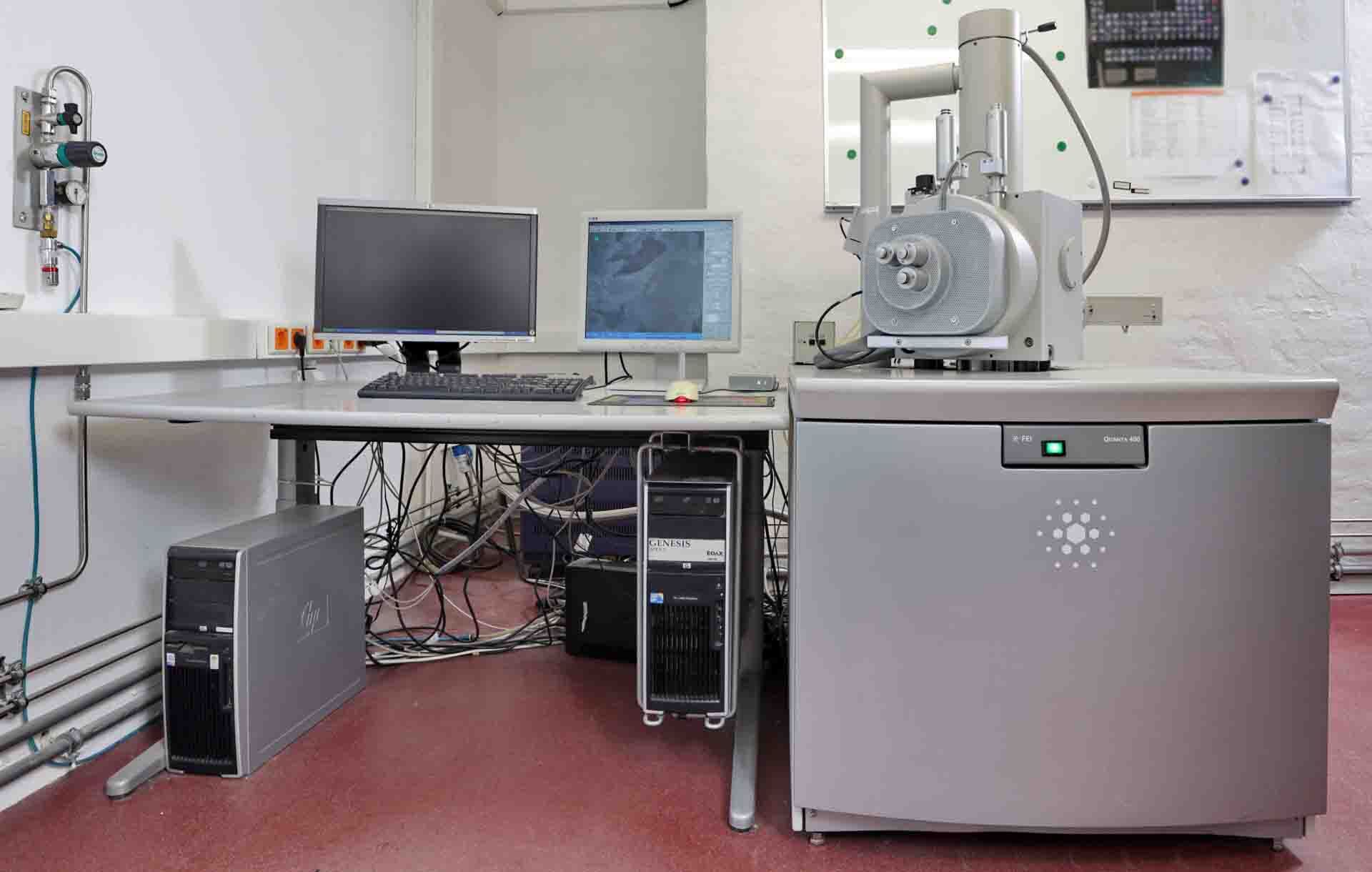
中古 PHILIPS / FEI Quanta 400 #293585905 を販売中
この商品は既に販売済みのようです。下記の同じようなプロダクトを点検するか、または私達に連絡すれば私達のベテランのチームはあなたのためのそれを見つけます。
タップしてズーム


販売された
ID: 293585905
Scanning Electron Microscope (SEM)
Dispersive X-Ray detector
Ultra-thin detector window
Backscatter electron detector
USB controller:
DC-Isolated USB 2.0 Interface
HV-VAC Controller:
DAC Uni / Bipolar: ± 10 V (8 x 16 bit)
ADC for emission (5 x 10 bit)
VAC
Operating voltages
(16) Digital inputs and outputs
Security link to vacuum system
Control power supply
Turn-on of power supply
Emergency turn-off of electronics at malfunction
Module for deflection coils:
(2) Single deflection systems
(4) Double deflection systems
Magnification: 4 step and 16 bit
Scan rotation
Scan shift fine
Correction of tilt
Orthogonality
Rotation offset
Module with bipolar power supplies:
DAC for current/voltage supplies (8 x 16 bit)
Beam shift
Tilt coils
Image shift coils
Stigmator coils (8 pole)
Filament imaging
Stigmator image
Power supply: 4 x ±500 mA, 4 x ±10 V
Module for objective lens and condenser lenses:
Unipolar power supply
Objective lens: 2 x 16 bit DAC (coarse, fine), maximum 6.5 A
(2) Condenser lenses: 16 bit DAC, maximum 6.5 A
Image system:
Maximum pixels: 16384 x 16384
Pixel clock: 200 ns
D/A Converter for analog input signals (4 x 12 bit)
Counter for mapping (12 x 16 bit)
Simultaneous acquisition
(4) Analogs
(12) Digital input signals
Image acquisition
Windows 2000: Up to 10 (x86, x64)
Full screen mode
Slow scan
Mapping
Oversampling for noiseless images: Up to 32000
Line averaging
Frame averaging
Reduced area scan
ROI Scan
Qualitative and quantitative with EDS/WDS
AVI Function
Signal monitor to control image signals
Trigger inputs and clock outputs for point
Mains synchronization for slow scan
Thumbnail bar for acquired images
Image processing:
Image browser
Loading and saving of images file types: TIFF, BMP, JPEG, PNG, GIF
Auto save function
Creation of image sections
Image rotation
Image labeling functions
SEM Parameters
Power supply:
Analog power supplies: ±15 V, +5 V, +12 V
Switching power supply for high power objective and condenser lenses
Module for PMTs: 2 x 0 to 1.5 kV
Module for scintillator: 12 kV
Grid voltage: 0 V to 400 V.
PHILIPS/FEI Quanta 400(略してQ400)は、材料特性評価および法医学分析に使用するために設計された走査型電子顕微鏡(SEM)である。SEMの中心にある小さな局所電子源である場の放出源を特徴としています。これにより、広範囲のサンプルタイプで高解像度の電子画像、地図、スペクトルを収集することができます。Q400は固体システムであるため、冷却に液体窒素を必要としません。このシステムは、サンプルに正確で正確なビーム電流を供給するために、大きなデジタル分解能コントローラを備えています。画像用には、最大0。5nmの解像度でハイコントラストの画像を提供することができ、表面トポグラフィの正確な決定を可能にします。さらに、SEM機能を使用して、EDS(エネルギー分散分光法)による化学マッピング情報を取得することができ、サンプル表面全体で素子マッピングを迅速に行うことができます。最適なSEMイメージングと解析を行うには、Q400に付属の「標準ダミー」サンプルホルダーを使用してサンプルを事前に準備する必要があります。これは、サンプルへの充電効果を低減または排除するために動作します。ラテックススフィアアタッチメントを使用してサーフェス間の相互作用を調べることができ、結晶方向はゴニオメーターを使用して収集することができます。サンプル表面の下では、Q400を使用して、一連の画像を収集することにより、より大きな領域の検索を通じて局所表面構造を探索することができます。これらをマージして縫い合わせることができます。このステッチ能力により、研究者は構造情報と組成情報のためにより広い領域を詳細に探索することができます。EDSマッピングと連動したSEM画像のキャプチャは、多くの質問を引き起こす可能性があります。例えば、標本の平均粒度、微細構造成分の大きさと分布、その他の形態学的または組成的分析。Q400は、これらの質問に答えることができる多くのソフトウェアプログラムをサポートしています。FEI Quanta 400は多目的で信頼性の高い走査型電子顕微鏡で、材料分析から法医学調査まで幅広い用途に使用できます。高解像度イメージング、EDSマッピング、ゴニオメーターアタッチメント、表面イメージング機能により、競合他社よりも優れています。
まだレビューはありません