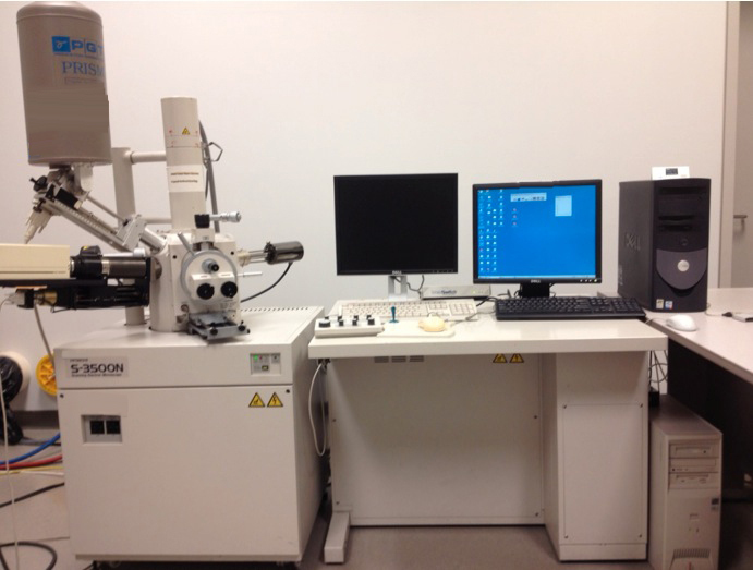
中古 HITACHI S-3500N #9046383 を販売中
この商品は既に販売済みのようです。下記の同じようなプロダクトを点検するか、または私達に連絡すれば私達のベテランのチームはあなたのためのそれを見つけます。
タップしてズーム


販売された
ID: 9046383
SEM
Resolution: 3.0nm at 25kV, secondary electron image
20nm at 3kV, secondary electron image
4.5nm at 25kV, backscattered electron image
Magnification: x15 - x300,000 (65 steps)
Accelerating voltage: 0.3 - 30kV
Variable pressure range: 1 - 270Pa
High and low vacuum operation
Backscatter electron detector
Energy Dispersive X-ray (EDS)
Orientation Imaging Microscopy (OIM)
Tungsten filaments
Optics:
Filament – pre-centered tungsten hairpin
Gun bias – self bias and continuously variable bias
Accelerating voltage: 10-30 kV
Emission current: 10-12 to 10-7
Gun alignment: 2-stage electromagnetic alignment
Condensers lens: 2-stage electromagnetic condenser
Objective lens: super conical lens
Objective lens aperture: 4 opening moveable aperture
Stigmator coil: 8-pole electromagnetic X/Y correction for astigmatism
Image shift: +/- 20 microns or more (for working distance 15 mm)
Specimen stage: Large-size eucentric stage
Movement range: 80mm x 40 mm
Tilt angle: 0° to +60°
Rotation angle: 360°
Image Display: Secondary electron
Scanning modes: TV scan, slow scan (4 steps), selected area scan, waveform, photo scan (4 steps), split screen and dual screen
Data display: accelerating voltage, magnification, micron scale, micron value, film number, working distance, value, time/date, photo magnification, detector
Data entry; input through full key board (alphabetic characters numerics and symbols)
Graphic input: straight lines, circles arrows etc.
Image memory:
Display (640 x480)
High resolution (1280 x 960)
Ultra-high resolution (2560 x 1920)
Computer system:
Hitachi soft-ware on Compaq PC with Windows 95, Pentium 160 MB processor
Hitachi software
Quartz PCI – imaging program
Auxiliary computer for EDS system – Dell, Intel Pentium
Two direct drive vacuum rotary vacuum pumps – Hitachi brand
Type VR16LPP 50 Hz, 60 Hz
Displacement 140, 160l/min
Ultimate pressure – 10-2 Pa
6W Infared Chamberscope
Components:
EDS system: PGT OPH045-1038
Backscattered detector
BSD system: Robinson
Computer system uses zip drives for downloading data, not USB
Original manuals and two Wehnelt cylinders.
HITACHI S-3500N Scanning Electron Microscope (SEM)は、多種多様な材料を高精度・高分解能で解析する強力な装置です。有機サンプルと無機サンプルの両方の詳細な画像を最大10万倍の倍率で生成することができます。この顕微鏡は、自動化されたナビゲーションシステムと低エネルギー環境チャンバーで設計されており、複雑なサンプルでも高解像度の簡素化と分析が可能です。電子光学系とエネルギー分散型X線検出器を内蔵したS-3500Nは、明るいフィールドと暗いフィールドの両方で比類のない精度とスキャン速度を提供します。Scanning Transmission Electron Microscopy (STEM;スキャン透過電子顕微鏡)機能により、軽い元素や構造物を簡単に検出できます。さらに、この顕微鏡は、最も複雑なナノ構造であっても、高コントラストで低ノイズの画像を取得することができます。HITACHI S-3500Nモデルは、マイナス染色、高真空、脱水、特殊気体大気においてもイメージングおよび分析を行うことができます。これは、サンプル内の様々なタイプのガスの制御導入を可能にする特別なガスインレット機能で構築されています。この機能は、検出器設計と組み合わせて、エピタキシャル層、絶縁体、半導体における微量元素または局所ドーパントに関する分析情報を生成することができます。S-3500Nは、直感的なグラフィカルユーザーインターフェイスを備えたユーザーフレンドリーなインストゥルメントです。組み込みアプリケーションスイートには、リアルタイムプロセス監視、画像測定、単粒子解析、スペクトルイメージング、egunセレクタなどのコンポーネントが含まれています。また、ユーザーはスコープのUSBインターフェースを介してさまざまな外部デバイスに接続することができ、複数のソースから画像とデータを同時に取得できます。HITACHI S-3500N走査型電子顕微鏡は、生体医学研究、半導体工学、微細加工、顕微鏡などの用途に最適です。その強力なイメージングと分析機能により、高品質で正確なデータと結果を求める人に理想的なツールとなります。
まだレビューはありません